Interview: Nicole Beaujean | N.B. text & advice
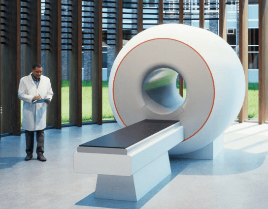
Parkinson's, ADHD, depression, autism. All brain disorders that we still do not know nearly enough about. 500 international researchers at the Donders Institute in Nijmegen deal daily with the many mysteries of our brain and the resulting behaviour. In about five or six years' time, they will get an important tool to assist them: the world's most powerful MRI scanner.
Anyone who has ever undergone an MRI scan may know that MRI stands for Magnetic Resonance Imaging. Such a scan is a medical examination with which a doctor takes detailed images of the inside of your body.
An MRI scanner works with a magnetic field, radio waves and a computer. A strong, superconducting magnet and radio waves are used to generate radio waves in the brain or another part of the body. An antenna captures the signals and the computer converts them into images.
More and faster
Prof David Norris, project leader and professor of MRI techniques at the Donders Institute: "We express the strength of the super magnet in Tesla, the unit for the field strength of a magnet. The field strength and thus power of a 3T magnet is two times that of the 1.5T. This allows significantly better images of the human body: more detailed and also much faster.
For medical diagnosis and research, an MRI scanner with a magnetic strength of 11.7 Tesla is already available in Paris. Seven Dutch research partners have now received a €19 million grant from the NWO (Netherlands Organisation for Scientific Research) for the world's first MRI scanner with a magnetic field strength of 14T."
The more tesla, the better?
About 98% of all investigations using an MRI scanner take place in a hospital; only 2% have a purely scientific purpose. The Donders Institute is one of the leading institutes for brain research; the largest in the Netherlands and one of the largest in Europe.
Norris: "So with us, it is not so much acute patients who end up in the scanner, but test subjects. We do research to gain more insight, not primarily to cure. With the 14T scanner, we can better visualise information exchange and brain activity and therefore take much more precise and faster measurements. This allows us to better diagnose diseases such as MS, cancer and 'small vessel disease'. MRI scans are also used for research on other organs: heart, prostate, liver.
The 14T super scanner is 13 times more sensitive than the 3T and three times more sensitive than the 7T," adds David Norris. "However, the total cost of the system is similar: around 25 million. So you could say that the 14T scanner is very good value for money."
Scientific community
The system will be assembled over the next few years by Philips, in collaboration with Tesla Engineering, Neoscan Solutions and Wavetronica. The 14T MRI scanner is expected to be placed on the Radboudumc site in 2027 or '28, but will be available to all scientists in the Netherlands.
"We will also collaborate with international partners," Norris said. "With this highly sensitive scanner, we want to enable new areas of research for the entire scientific community. We therefore cordially invite our colleagues worldwide to submit their research proposals."

Consortium
The consortium of seven partners is led by Radboud University's Donders Institute for Brain, Cognition and Behaviour.
The other six partners of the consortium are: Amsterdam UMC, Leiden University Medical Centre (LUMC), Maastricht University, Radboudumc, Spinoza Centre for Neuroimaging - KNAW and University Medical Centre Utrecht (UMCU).